Elbow Dysplasia
What is Elbow Dysplasia?
Elbow dysplasia is the most common cause of lameness on a front leg in young dogs, especially of the larger breeds. Dysplasia refers to abnormal development, in this case of the elbow joint.
The elbow is formed from where three bones meet: the humerus, which is the upper limb from shoulder to elbow; the ulna, from the elbow to the paw along the back of the limb; and the radius, which supports the major weight-bearing along the front of the lower limb. All three of these bones need to grow and develop normally and at the same rate so that they fit perfectly together. If there are any abnormalities along these lines, or if the cartilage lining the elbow joint does not form properly, then dysplasia or an abnormal formation results.
Elbow dysplasia can take several different forms. They can be seen individually or combined. While all of the variations are distinct and probably develop in different ways, they all produce loose pieces of bone and/or cartilage within the joint that act as irritants, much as a pebble does in your shoe. These variations also are primary problems that invariably lead to arthritis in the elbow.
Traditionally, there are four conditions that result from abnormal development of the elbow:
- Medial coronoid disease
- Osteochondritis dissecans (OCD)
- Ununited anconeal process (UAP)
- Elbow incongruity
These conditions may occur in isolation or in combination and affect the way the bones fit together and/or the quality of the joint cartilage. This ultimately results in elbow osteoarthritis and forelimb lameness (limping). The degree of osteoarthritis that a patient develops is variable and often unpredictable. Likewise, the degree to which the osteoarthritis affects an individual patient is often unpredictable. Unfortunately, osteoarthritis in any joint in the body is incurable, but with appropriate care, a good quality of life can be attained for your pet.
Medial coronoid disease
Concentration of forces in the medial compartment of the elbow joint can result in stress fractures forming within a peninsula of bone called the medial coronoid process of the ulna. These stress fractures can result in fragments forming within the joint (fragmented coronoid process). This ultimately leads to inflammation within the elbow (osteoarthritis).
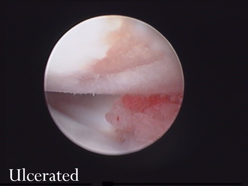
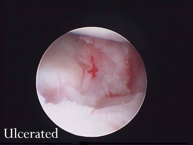
That part of the elbow where the medial coronoid process and humerus come into contact is called the medial compartment. Commonly, there is severe wear and loss of cartilage in the medial compartment resulting in “bone on bone” contact. This condition is often referred to as medial compartment disease.
Osteochondritis dissecans (OCD)
Osteochondrosis occurs as a result of a problem with the way bone and cartilage form in a joint. In the elbow, this occurs in the medial trochlear ridge of the humerus. A flap of cartilage can peel away from the humerus resulting in osteochondritis dissecans (OCD), a painful condition resulting in osteoarthritis.
Ununited anconeal process (UAP)
Occasionally, a triangular process of bone and cartilage called the anconeal process doesn’t attach properly to the top part of the ulna during development. This loose piece of bone and cartilage results in discomfort and osteoarthritis.
Elbow incongruity
At times, the bones in the elbow don’t fit together properly. This can be as a result of a radius or ulna that is too short, or, as a result of an ulnar notch that is the wrong shape. These problems are thought to contribute to the development of medial compartment disease, fragmentation of the coronoid process, UAP and the resultant osteoarthritis.
Symptoms
The first sign of elbow dysplasia is a mild to moderate front leg lameness in a young dog between 4 to 10 months. If the problem is not diagnosed at this stage, more lameness may be seen as severe arthritis sets in. It is mostly large breed dogs that suffer from elbow dysplasia, but there are small dogs that can be affected also.
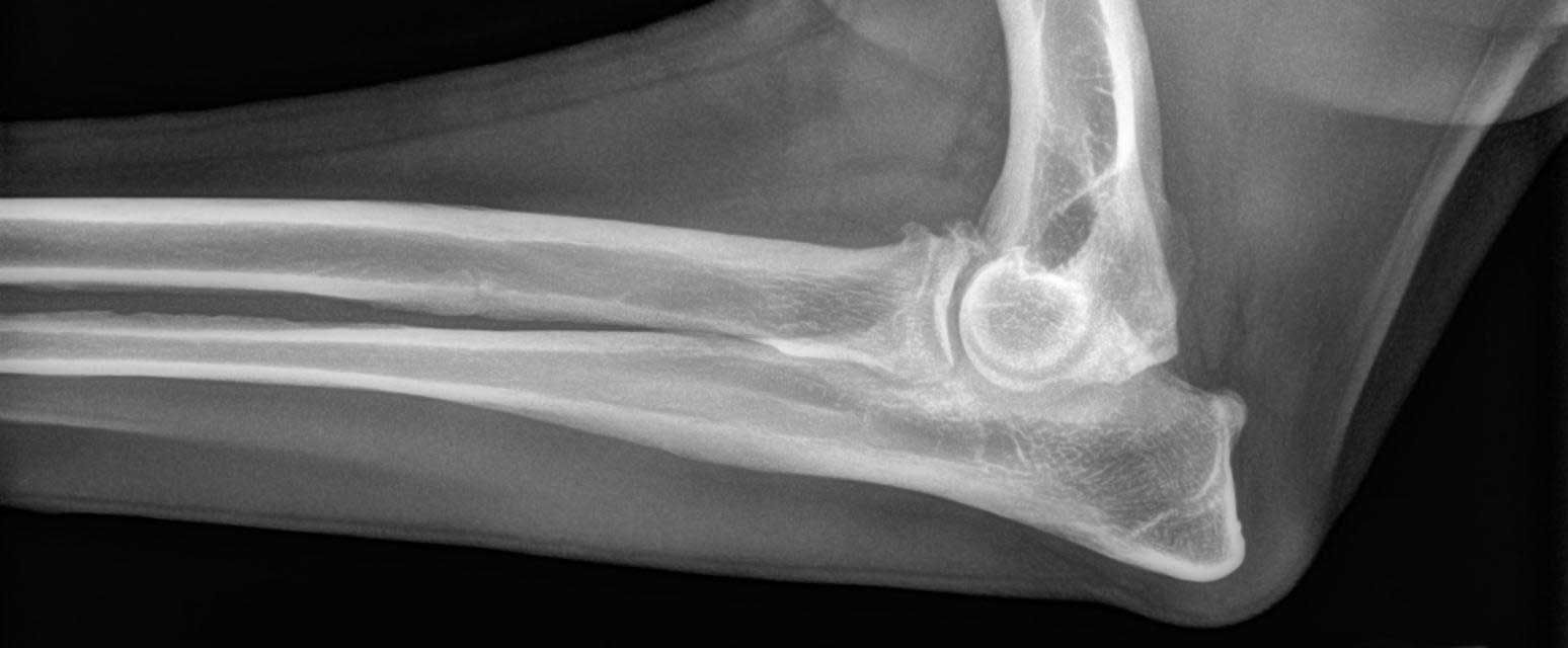
Diagnosis
The history of front leg lameness in a young, large breed dog is suggestive of elbow dysplasia. When examined, the elbow may show pain, thickening or swelling, and restricted movement. Radiographs of the elbow will usually confirm the diagnosis. There are usually radiographic signs that suggest the diagnosis. Advanced imaging studies, particularly computed tomography (CT scan), may be helpful.
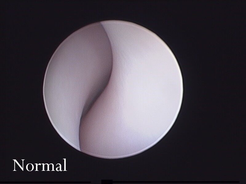
Ultimately, surgery may be needed to provide a complete diagnosis. In recent years, such exploration is most often done with an arthroscope (a camera that is inserted into the joint without the need of major open surgery).
Treatment
Surgical intervention by an orthopaedic specialist provides the preferred means of treatment for many cases of elbow dysplasia. There are numerous specialists that we can refer you to for treatment but they are all located in the Brisbane area. There is a specialist who travels to Rockhampton about every 6 weeks that may work in some instances. However, unfortunately a diagnosis of elbow dysplasia needs treatment as soon as possible to achieve the best outcome. We recommend a specialist appointment within one week of diagnosis.
Surgical Options
Arthroscopic fragment removal and subtotal coronoidectomy – MOST COMMON AND SUCCESSFUL
This treatment strategy involves using an arthroscopy to guide the removal of fragments of cartilage/bone from the elbow joint or, the majority of the medial coronoid process. This procedure is performed minimally invasively, with the majority of dogs walking and able to be discharged the day after surgery. Removal of the OCD cartilage flap is also achieved using arthroscopy.
Proximal abducting ulnar osteotomy (PAUL)
This technique involves performing a cut in the ulna and stabilising it using a special titanium plate and screws. The aim is to reduce the load that is taken by the diseased part of the elbow joint resulting in a more comfortable joint.
Dynamic ulnar osteotomy
Sometimes, a type of ulnar osteotomy is performed that is not stabilised using a plate. These techniques are often used if there is joint incongruity present, that is, if the radius and ulna don’t align with the humerus properly. They allow the bone to move into a more congruent position such that the bones fit together better. Combined with an accurately placed screw, an ulnar osteotomy may specifically benefit patients with UAP.
Non-Surgical Management
Some dogs with elbow dysplasia may be determined to be best treated with non-surgical therapies. These usually include weight management, exercise adjustment, physiotherapy and supplements that may improve joint health.
Prognosis
The aim of treatment for dogs with elbow dysplasia, is to improve their comfort level. Unfortunately, it is impossible to cure the disease and the secondary osteoarthritis that results. However, with an appropriate assessment by the speciailist surgeons, a management regime can be tailored to achieve the best possible outcome for your pet.